March 19th - Pregnant Dolphin Stranded in Hilton Head
- Nicole Principe
- Mar 20, 2024
- 2 min read
Updated: Apr 11, 2024
On the afternoon of March 19th, just as our team finished up a necropsy on a dolphin recovered in Kiawah, we were notified of another deceased dolphin near Sea Pines in Hilton Head. First responders onsite were able to move the dolphin above the high tide line so it would be secure overnight.

The next morning, our partners with NOAA's Coastal Marine Mammal Assessments Program, drove down to retrieve the dolphin and transport it back to our necropsy lab at Hollings Marine Lab. Initial observations of this adult female bottlenose dolphin suggested she was likely pregnant. She was very robust and was leaking a significant amount of milk from her mammary glands.

Morphometric data was collected and a necropsy was initiated on the afternoon of March 20th by Lauren Rust, Nicole Principe, Bonnie Ertel, and Colin Perkins-Taylor.
Upon opening up the abdominal cavity, it was immediately clear this animal was pregnant with a near full-term fetus. However, instead of the fetus being found in the uterus, it was lying on top of the intestines. The fetus had ruptured from the uterus and likely died before causing infection and maternal death.

It is unclear what caused the fetus to rupture from the uterus, but additional samples were collected from both animals for Brucella testing. It is undoubtedly sad to lose both a reproducing female and an unborn calf, but hopefully test results will provide more answers.
Dolphin Pregnancy and Birth
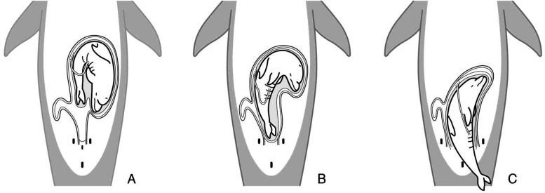
Peak mating season for bottlenose dolphins typically occurs during the spring. Gestation lasts a full 12 months with peak birthing season also occurring in the spring. Dolphin calves are usually folded within the uterus and are born tail first (breech position) so they do not drown during the birthing process. They are born darker in color and still show evidence of folds from being in the womb.
One study by Terasawa et al. (2021) used ultrasonography to estimate how fetal position would be within the birth canal and how the fluke will emerge first from the mother's body at parturition. In our case, although the fetus ruptured from the uterus, it appeared to be in the correct orientation for birth.

Terasawa, F., Akiyama, H., Sakuragi, T., Haneda, S., & Shirakata, C. (2021). Embryo and fetal position during pregnancy by ultrasonographic examinations in bottlenose dolphin (Tursiops truncatus). The Journal of Veterinary Medical Science, 83(7), 1138–1143. https://doi.org/10.1292/jvms.21-0181
Comments